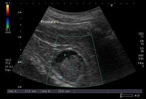
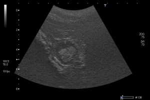
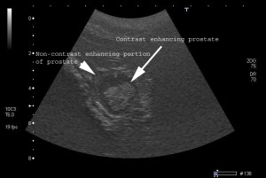
Last week’s ground breaking prostate study using targeted ultrasound contrast yielded some exciting results. The post-contrast images were incredible and revealed after five minutes that the abnormal appearing prostatic core was contrast-enhancing, while the peripheral prostatic tissue, particularly on the right, did not demonstrate enhancement. These initial results demonstrate potential promise in targeted ultrasound contrast’s ability to differentiate normal from cancerous portions of the prostate.
Following the procedure the patient woke up from anesthesia uneventfully. No side effects to the contrast were observed during monitoring of the heart rate, respiratory rate, or ECG.
Conclusions from Images:
1) Contrast-enhancing prostatic core. This finding suggests abnormal (potentially cancerous) tissue.
2) Non-contrast enhancing prostatic margin. This finding suggests non-neoplastic tissue.
3) No sublumbar lymph node enlargement.
Biopsy Results:
A biopsy using an 18 gauge spring loaded needle with a 2 cm core was performed of both the non-enhancing and the enhancing tissue. Results are pending.
Implications:
The implications of this study may be far-reaching, allowing for early detection of prostate cancer in both dogs and people using non-invasive ultrasound coupled with targeted ultrasound contrast. Historically, medical testing and imaging were initially performed on healthy small animals such as rats and mice during clinical trials, with findings later applied to humans. However, a new clinical research philosophy is beginning to emerge where cutting edge diagnostics and medical care are being offered to both humans and pets afflicted with similar conditions. This comparative testing model may offer a better understanding of cancer in both humans and animals.


