“What’s Your Diagnosis?” Challenge Winner & Answer: Jan-Mar 2012
Special thanks to Dr. Henry Yoo of Crenshaw Animal Hospital in Torrance, CA for sending us this case.
Case History
The patient is a 7-year-old, 75-pound M/C Lab who lost over 10 lbs since August. Vomiting occasionally and very low appetite.
Survey Radiographs
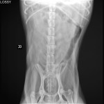
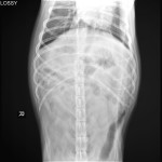
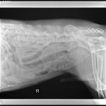
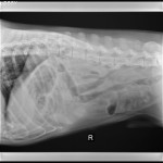
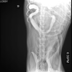
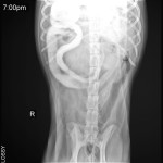
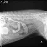
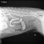
Four survey radiographs of the abdomen are available for interpretation. (Click on each to view larger images.)
Barium Series
A barium series was also performed. A few representative images from the barium series are included. (Click on each to view larger images.)
WYD Answer
Interpretation: There is a moderately dilated segment of small intestine present within the right mid-abdomen. Along the cranial margin of this dilated segment of small intestine there is a sharply marginated, almond shaped, peripherally soft tissue opaque, centrally fat opaque structure that is likely present within the lumen. The structure is best seen on the lateral views. The remaining segments of small intestine are normal in diameter. Abdominal detail is normal.
Conclusion: The segmental small intestinal dilation is consistent with a foreign body obstruction. The structure within the bowel lumen is characteristic of a peach pit.
Outcome: This dog went to surgery and a peach pit causing an intestinal obstruction was removed. The dog recovered uneventfully from surgery and was discharged the following day.
Congratulations to Grant Mayne, a San Diego relief vet, for winning this challenge!


